Early mammals were small, with small brains, an emphasis on olfaction, and little neocortex. Neocortex was transformed from the single layer of output pyramidal neurons of the dorsal cortex of earlier ancestors to the six layers of all present-day mammals. This small cap of neocortex was divided into 20-25 cortical areas, including primary and some of the secondary sensory areas that characterize neocortex in nearly all mammals today. Early placental mammals had a corpus callosum connecting the neocortex of the two hemispheres, a primary motor area, M1, and perhaps one or more premotor areas. One line of evolution, Euarchontoglires, led to present-day primates, tree shrews, flying lemurs, rodents, and rabbits. Early primates evolved from small-brained, nocturnal, insect-eating mammals with an expanded region of temporal visual cortex.
The agranular area of the PFC led to the capacity to internally model the environment and plan future moves. As anthropoids emerged as diurnal primates, the visual system specialized for detailed foveal vision. Other adaptations included an expansion of prefrontal cortex and insular cortex. The human and chimpanzee-bonobo lineages diverged some 6-8 million years ago with brains that were about one third the size of modern humans. Over the last 2 million years, the brains of our more recent ancestors increased greatly in size, especially in the prefrontal, posterior parietal, lateral temporal, and insular regions. .
Specialization of the two cerebral hemispheres for related, but different functions became pronounced, and language and other impressive cognitive abilities emerged. The emergence of the granular area of the PFC allowed simians to model their own thoughts. This led to the ability to form future scenarious and outlooks - imagination. This was the slowest of mental skills but was the most far reaching. This led to such things like long term planning and Machavellian plots. The human capacity for this outshone all other life forms.
The human brain size is limited by the size of the skull. This is limited, not by itself but by females to give birth to young that have critically large heads. Many animals have increased their cognition by scaling up the number of neurons. More neurons – more connections and problem solving abilities. The prefrontal cortex solved this problem by growing a thin sheet on top of the existing brain matter. This had to be tucked in to available spaces and caused the folds so easily apparent.
AI has its own scaling problem, that of the size of chips and the failure of Moore’s Law. Owing to the quantum nature artificial neurons are effected by close electron interference and noise failure.
Richard Sutton invented the temporal difference learning to overcome the problem of how to credit good actions with bad actions in a sequence of events. One action may be a winning one but which one. It could have been any any or more than one in a sequence. How to reward the actual winners was a complicated decision/s. He thought of using a critic to predict possible good actions and an actor to carry out these actions. If both were positive then a reward of some sort was instigated. This was not a single action but was repeated multiple times to find the most probable outlook.
It was originally thought that the brain rewards with dopamine when a positive action is effected. It was Dayan and Montague found a decade later that this was not the case. They found that predictive rewards were the catalyst for dopamine release and if the action was expected then there was no release. Also the closer in time between prediction and fulfilment released more dopamine.
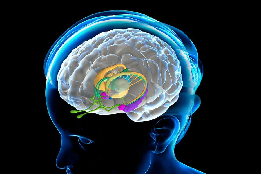
The anatomy of the basal ganglia is complex since it is spread throughout the forebrain. Its components can be divided into input nuclei, output nuclei and intrinsic nuclei. Input nuclei receive information, which is then relayed to intrinsic nuclei for processing, and further passed to output nuclei:
The basal ganglia are a group of structures near the center of your brain that form important connections. These connections allow different areas of the brain to work together. The basal ganglia manage the signals your brain sends that help you move your muscles.
The basal ganglia are best known for how they help your brain control your body’s movements. However, ongoing research continues to uncover other ways that the basal ganglia interact with other parts of the brain. Though experts continue to uncover more about the inner workings of the basal ganglia, there’s much about them that remains unknown.
Input Nuclei: Caudate nucleus and putamen (neostriatum).
Intrinsic Nuclei: External globus pallidus, Subthalamic nucleus, Pars compacta of the substantia nigra.
Output Nuclei:Internal globus pallidus, Pars reticulata of the substantia nigra.
The basal ganglia are a key part of the network of brain cells and nerves that control the body’s voluntary movements. They can approve or reject movement signals that your brain sends, filtering out unnecessary or incorrect signals. This lets it control certain muscles without also using other muscles that are nearby. If the basal ganglia approve a signal, it continues to the motor pathways, the nerves that eventually carry the signal down the spinal cord and nerves to their destination muscle. If they don’t approve the signal, they redirect it into an area where other brain cells dampen those signals until they stop. The parts of your brain that process information from your senses, namely sight, sound, smell, taste and touch, also send that information to your basal ganglia. That sensory information helps the basal ganglia refine your movements further.
Another job of the basal ganglia is processing how you evaluate goals and risks. It also processes signals that affect your emotions and your motivation. That means it also plays a role in learning and forming habits, planning and carrying out tasks, and more.
The hippocampus is a vital part of the human brain, located within the medial temporal lobe. Its primary roles include:
Memory formation : The hippocampus is crucial for converting short-term memories into long-term memories. It helps us remember facts and events.
Spatial Navigation: It plays a key role in helping us navigate our environment and remember spatial relationships.
Emotional Regulation: The hippocampus is involved in regulating emotions by interacting with other parts of the brain, particularly the amygdala.
Damage to the hippocampus can lead to difficulties in forming new memories (anterograde amnesia) or recalling past memories (retrograde amnesia).
Humans are very sight orientated animals and much of the virtual scenarios arise from the eyes. Visual data from the retinas of the eyes travel to the visual cortex via the thalamus, where it synapses in a nucleus known as the lateral geniculate. This information is then passed from the lateral geniculate to V1, the very first region of the visual cortex. V1 is also called the primary visual cortex. The stimuli passes though V2-V6 and the images is improved and ready to pass to other parts of the brain.